Presenter Name: Taylor Otterness
Description
Purpose:
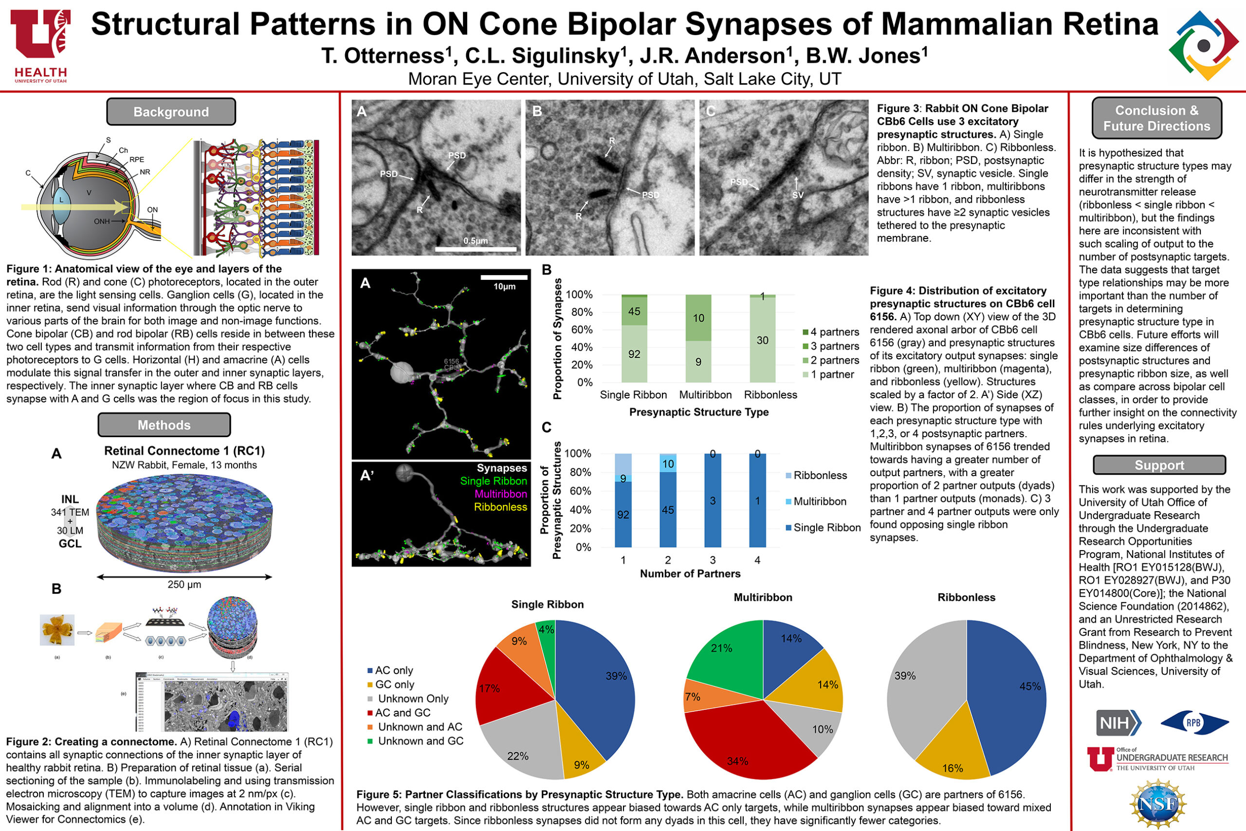
Connectivity within the nervous system is precise and disruptions lead to disease, yet the rules governing this connectivity remain unknown. Recent efforts have shown that different types of ON cone bipolar cells in the neural retina show preferences in the selection and frequency of presynaptic structure types used for signal transmission. However, it is not yet known if these differences are related to the quantity and type of postsynaptic partner.The goal of this study was to analyze the synaptic output of CBb6 cells, a type of ON cone bipolar cell with diverse presynaptic structure types to identify patterns in how ON cone bipolar cells interact
with their postsynaptic partners. Methods:
The rabbit connectome dataset (RC1) used in the study was sampled from a 0.25mm diameter patch of mid-peripheral retina from a healthy 13 month old female Dutch-Belted rabbit and serially sectioned at 70 nm. The resulting sections were imaged at ultrastructural resolution (2nm/px) using transmission electron microscopy. Postsynaptic partners of CBb6 cell 6156's bipolar-specific presynaptic structures (multi-ribbons, single ribbons, and ribbonless synapses) were annotated to classification using the Viking Viewer for Connectomics. Statistical analyses were conducted in Microsoft Excel and investigated further with 3D rendering and graph visualization of connectivity. The factors tracked for comparison were presynaptic structure type, target number, and postsynaptic partner type. Results:
Although the ribbonless presynapses of 6156 were restricted to single postsynaptic partners, both single and multiribbon presynapses showed one or two partners, but only single ribbons exhibited more than two postsynaptic partners, despite some multiribbon synapses containing up to 6 ribbon structures. As the different presynaptic structure types are thought to differ in neurotransmitter release (ribbonless < single ribbon < multiribbon), these findings are inconsistent with scaling of presynaptic structure type with the number of postsynaptic targets. Preliminary partner classification reveals that amacrine and ganglion cells were both found postsynaptic to all presynaptic structure types and some cells received input from 6156 by multiple presynaptic structure types. More detailed analyses of partner type are ongoing. Conclusion:
The preliminary findings from this study suggest that the presynaptic structure type may not correlate with either the number or type of postsynaptic targets for this particular cell type. Ongoing efforts aim to further classify the partner cells and analyze more CBb6 cells. Future efforts will look at ribbon size and cumulative postsynaptic area to determine if those measures are better predictors. If and how these findings extend across other ON cone bipolar cell types will be important for understanding why these cell types differentially utilize these presynaptic structure types and together will inform our understanding of the connectivity rules driving synapse formation in the retina and possibly the brain.
University / Institution: University of Utah
Type: Poster
Format: In Person
Presentation #D16
SESSION D (3:30-5:00PM)
Area of Research: Health & Medicine
Email: u0907150@utah.edu
Faculty Mentor: Crystal Sigulinsky